Category Archives: Biofilm

Research – Researchers identify biofilm hotspots, document danger zones

Food Safety News

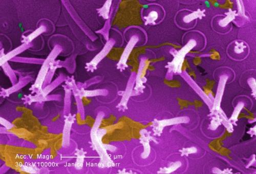
Scientists in Austria have investigated where biofilms hide and what bacteria can be found in them.

Biofilms are proven sources of contamination in the food industry. They can cause additional costs in production and can be a danger to consumer health.

Researchers at the unit of food microbiology at Vetmeduni Vienna looked at biofilms in an Austrian meat processing environment that included pork, poultry and beef. Knowledge gained on presence and composition, published in the International Journal of Food Microbiology, could help to prevent and reduce biofilm formation within food processing environments.

Research – Biofilm-forming ability of pathogenic strains isolated from retail food in Poland

Journal of Food Protection

Biofilms have a significant impact on food safety in the food industry. Many foodborne outbreaks have been associated with pathogenic strains that can form a biofilm. The present study aimed to examine the ability to form biofilms by pathogenic strains collected from retail food samples under the Official Control and Monitoring Program in Poland. Biofilm formation was assessed by the qualitative detection of EPS production on Congo Red Agar, tube method, crystal violet biofilm assay, and MTT assay. A total of 40 isolates from food samples (10 strains for each of the species Listeria monocytogenes, Staphylococcus aureus, Escherichia coli, and Bacillus cereus ) were examined. . . The study classifies strains as adherent strain, slightly adherent, nonadherent (A, SA and N); as weak, moderate, and strong biofilm formation (WBF, MBF and SBF); and weak, moderate, and high metabolic activity (WMA, MMA and HMA). Incubation conditions and time influenced the biofilm levels formed. Moreover, growth medium had a significant impact on biofilm formation. Results showed that 22.5% strains demonstrated A type in LBB and 77.5% SA type in LBB in the test tube while the stronger adhesion was obtained in BHI with 2% sucrose. Among the isolates incubated in the BHI medium with 2% sucrose, A type was observed in 60% of isolates (60%).. CV assay result show that the after 24 h incubation in LBB, SBP was 7,5% while after 48 h – 37.5% tested strains. For BHI medium supplemented with 2% sucrose after 24 h incubation strains 42.5% was classified as SBP and 37.6% after 48h.MTT assay result indicate that 15% strains incubate in LBB (24h) was HMA, and after 48h incubation time HMA show 25%. For BHI medium supplemented with 2% sucrose after 24 h or 48 h incubation strains 70% and 85 % was classified as HMA.

Research – Washing away stubborn biofilms using fungal cleaning products

Science Daily biofilm

Lurking inside pipes and on the surfaces of indwelling medical devices, slimy layers of bacteria, called biofilms, cause problems ranging from largescale product contamination to potentially fatal chronic infections. Biofilms are notoriously difficult to eliminate — not surprising given that one of their main functions is to protect encased bacteria from threats such as predation, antibiotics, and chemical cleaning agents.

Bleach, harsh oxidizing cleaning products, and petrochemical-derived detergents called surfactants combined with scrubbing are the most effective methods of removing biofilms. However, bleach and harsh chemicals are obviously unsuitable for use in biological settings, and while surfactants are used in products such as hand soap and cosmetics, many are toxic to the environment and can damage the surfaces that they are used on.

But in a study published this month in peer-reviewed journal Langmuir, researchers from the University of Tsukuba have found a new way of tackling biofilms, using cleaning agents derived from microbes themselves.

Research – Effect of essential oils on pathogenic and biofilm-forming Vibrio parahaemolyticus strains

Tandfonline

In this study, the effect of three essential oils (EOs) – clove oil (CO), thyme oil (TO), and garlic oil (GO), which are generally recognized as safe – on the planktonic growth, minimum inhibitory concentration (MIC), minimum bactericidal concentration (MBC), motility, biofilm formation, and quorum sensing (QS) of Vibrio parahaemolyticus was investigated. All three EOs showed bacteriostatic activity, with MICs in the range 0.02%–0.09% (v/v). CO and TO completely controlled planktonic growth at 0.28% and 0.08% (v/v), which is four times their MIC (4 × MIC), after 10 min, whereas GO completely controlled growth at 0.36% (v/v) (4 × MIC) after treatment for 20 min. V. parahaemolyticus motility was significantly reduced by all three EOs at 4 × MIC (0.28% for CO, 0.08% for TO, and 0.36% for GO), whereas QS was controlled and biofilm formation reduced by all three EOs at 8 × MIC (0.56% for CO, 0.16% for TO, and 0.72% for GO) after 30 min of treatment. These results suggest that CO, TO, and GO have a significant inhibitory effect on V. parahaemolyticus cells in biofilm sand thus represent a promising strategy for improving food safety. These results provide the evidence required to encourage further research into the practical use of the proposed EOs in food preparation processes.

Research – Escherichia coli Biofilms

Springer Link ecoli

Escherichia coli is a predominant species among facultative anaerobic bacteria of the gastrointestinal tract. Both its frequent community lifestyle and the availability of a wide array of genetic tools contributed to establish E. coli as a relevant model organism for the study of surface colonization. Several key factors, including different extracellular appendages, are implicated in E. coli surface colonization and their expression and activity are finely regulated, both in space and time, to ensure productive events leading to mature biofilm formation. This chapter will present known molecular mechanisms underlying biofilm development in both commensal and pathogenic E. coli.

Research – Mom was right about not touching slime; researchers looking at pathogens in food facilities

Food Safety News biofilm

New research underway seeks to determine how and where bacteria hide in food processing plants so that they can be eliminated, to the benefit of public health and the bottom line of food companies.

The project involves scientists at Texas A&M, Stanford University, and the USDA’s Agricultural Research Service U.S. Meat Animal Research Center’s pilot meat processing facility in Clay Center, NE. Their work has the backing of a $479,000 grant from the U.S. Department of Agriculture (USDA).

While the project could ultimately benefit food facilities from fresh produce processing plants to candy makers, the researchers will be taking an extra close look at the meat industry.

Led by Sapna Chitlapilly Dass, a faculty member in Texas A&M’s Department of Animal Science, the team will be looking at “hotspots” that easily harbor biofilms, also known as slime. Dass and the other researchers are trying to figure out not only where bacteria like Salmonella, Listeria, and E. coli O157: H7 are hiding, but what sanitizers they have become resistant to.

Research – Effectiveness and Functional Mechanism of a Multicomponent Sanitizer against Biofilms Formed by Escherichia coli O157:H7 and Five Salmonella Serotypes Prevalent in the Meat Industry

Journal of Food Protection

ABSTRACT

Biofilm formation by Escherichia coli O157:H7 and Salmonella enterica at meat processing plants poses a potential risk of meat product contamination. Many common sanitizers are unable to completely eradicate biofilms formed by these foodborne pathogens because of the three-dimensional biofilm structure and the presence of bacterial extracellular polymeric substances (EPSs). A novel multifaceted approach combining multiple chemical reagents with various functional mechanisms was used to enhance the effectiveness of biofilm control. We tested a multicomponent sanitizer consisting of a quaternary ammonium compound (QAC), hydrogen peroxide, and the accelerator diacetin for its effectiveness in inactivating and removing Escherichia coli O157:H7 and Salmonella enterica biofilms under meat processing conditions. E. coli O157:H7 and Salmonella biofilms on common contact surfaces were treated with 10, 20, or 100% concentrations of the multicomponent sanitizer solution for 10 min, 1 h, or 6 h, and log reductions in biofilm mass were measured. Scanning electron microscopy (SEM) was used to directly observe the effect of sanitizer treatment on biofilm removal and bacterial morphology. After treatment with the multicomponent sanitizer, viable E. coli O157:H7 and Salmonella biofilm cells were below the limit of detection, and the prevalence of both pathogens was low. After treatment with a QAC-based control sanitizer, surviving bacterial cells were countable, and pathogen prevalence was higher. SEM analysis of water-treated control samples revealed the three-dimensional biofilm structure with a strong EPS matrix connecting bacteria and the contact surface. Treatment with 20% multicomponent sanitizer for 10 min significantly reduced biofilm mass and weakened the EPS connection. The majority of the bacterial cells had altered morphology and compromised membrane integrity. Treatment with 100% multicomponent sanitizer for 10 min dissolved the EPS matrix, and no intact biofilm structure was observed; instead, scattered clusters of bacterial aggregates were detected, indicating the loss of cell viability and biofilm removal. These results indicate that the multicomponent sanitizer is effective, even after short exposure with dilute concentrations, against E. coli O157:H7 and S. enterica biofilms.

HIGHLIGHTS
  • No viable biofilm cells were detected after treatment with the multicomponent sanitizer.
  • Prevalence of both pathogens was low after treatment with the multicomponent sanitizer.
  • SEM analysis revealed that treatment dissolved the EPS matrix and destroyed the biofilm.

Research – Cholera studies reveal mechanisms of biofilm formation and hyperinfectivity

UCSC

Free-swimming cholera bacteria are much less infectious than bacteria in biofilms, aggregates of bacterial cells embedded in a sticky matrix that form on surfaces. This accounts for the surprising effectiveness of filtering water through cloth, such as a folded sari, which can reduce infections dramatically in places where the disease is endemic, despite the fact that individual cholera bacteria easily pass through such a filter.

A new study led by researchers at UC Santa Cruz goes a long way toward explaining the hyperinfectivity of cholera biofilms. The study, published the week of April 20 in the Proceedings of the National Academy of Sciences (PNAS), is one of several new papers on cholera biofilms from the laboratory of UCSC microbiologist Fitnat Yildiz.

Research – Bacteria form biofilms like settlers form cities

Science Daily biofilm

Biofilms are composed of bacteria living in a densely packed and organized community. Research paired high-level imaging tools with an algorithm to track a biofilm as it formed. Biofilm growth at multiple scales, they found, mimics aspects of how cities emerge from individual settlers.

The findings show that, as individual bacteria multiply and grow into a dense and sticky biofilm, such as the community that forms dental plaque, their growth patterns and dynamics mirror those seen in the growth of cities.

“We take this ‘satellite-level’ view, following hundreds of bacteria distributed on a surface from their initial colonization to biofilm formation,” says Hyun (Michel) Koo, a professor in Penn’s School of Dental Medicine and senior author on the work. “And what we see is that, remarkably, the spatial and structural features of their growth are analogous to what we see in urbanization.”

This new perspective on how biofilms grow could help inform efforts to either promote the growth of beneficial microbes or break up and kill undesirable biofilms with therapeutics.

Research – Biofilm formation by South African non-O157 Shiga toxigenic Escherichia coli on stainless steel coupons

NRC

This study examined the biofilm-forming ability of six non-O157 Shiga-toxin-producing Escherichia coli (STEC) strains: O116:H21, wzx-Onovel5:H19, O129:H21, O129:H23, O26:H11, and O154:H10 on stainless steel coupons after 24, 48, and 72 h of incubation at 22 °C and after 168 h at 10 °C. The results of crystal violet staining revealed that strains O129:H23 and O154:H10 were able to form biofilms on both the submerged surface and the air–liquid interface of coupons, whereas strains O116:H21, wzx-Onovel5:H19, O129:H21, and O26:H11 formed biofilm only at the air–liquid interface. Viable cell counts and scanning electron microscopy showed that biofilm formation increased (p < 0.05) over time. The biofilm-forming ability of non-O157 STEC was strongest (p < 0.05) at 22 °C after 48 h of incubation. The strongest biofilm former regardless of temperature was O129:H23. Generally, at 10 °C, weak to no biofilm was observed for isolates O154:H10, O116:H21, wzx-Onovel5:H19, O26:H11, and O129:H21 after 168 h. This study found that temperature affected the biofilm-forming ability of non-O157 STEC strains. Overall, our data indicate a high potential for biofilm formation by the isolates at 22 °C, suggesting that non-O157 STEC strains could colonize stainless steel within food-processing facilities. This could serve as a potential source of adulteration and promote the dissemination of these potential pathogens in food.